
Thermography Technique in Diagnosing Equine Musculoskeletal Diseases:
A Comprehensive and Specialized Overview
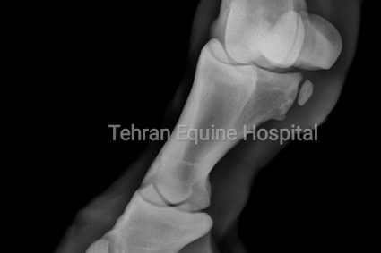
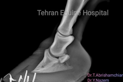
Thermography, or thermal imaging, is a non-invasive and safe technique widely used in veterinary medicine for diagnosing diseases and issues in the musculoskeletal system of horses. This technique works by measuring and displaying changes in the surface temperature of the body. Using infrared-sensitive cameras, it creates a thermal map of different areas of the body. Thermography plays a crucial role in early disease detection and the identification of hidden issues, as it can help detect inflammation and tissue damage before clinical signs become visible.
Principles of Thermography:
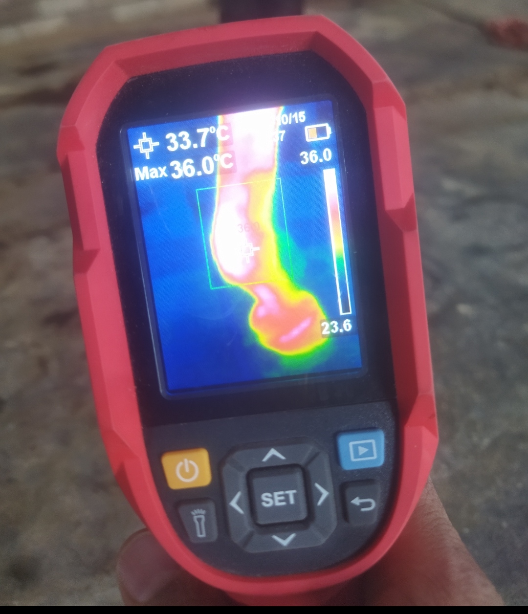
Thermography operates on the principle that inflammation or tissue damage leads to increased blood flow, which in turn raises the temperature in the affected area. Thermographic cameras are capable of detecting and recording these temperature changes, producing color thermal images where warmer areas are displayed in brighter colors (typically red or yellow) and cooler areas in darker shades (blue or green).
Applications of Thermography in Diagnosing Equine Musculoskeletal Diseases:
1. Early Detection of Inflammation and Soft Tissue Injuries: Thermography can identify inflammation and microscopic tissue damage before clinical signs appear. This enables veterinarians to intervene early, preventing further disease progression.
2. Assessment of Tendon and Ligament Injuries: Tendon and ligament injuries are common in athletic horses. Thermography can detect these injuries by identifying areas with elevated temperature in tendons and ligaments.
3. Evaluation of Arthritis and Joint Inflammations: Inflamed joints have increased blood flow, which produces more heat. Thermography can assist in diagnosing arthritis and other joint issues, providing accurate information for treatment decisions.
4. Monitoring Recovery Post-Treatment: Thermography can be used to evaluate the effectiveness of treatments and monitor recovery progress. This technique allows veterinarians to assess improvement or recurrence of disease by comparing thermal images before and after treatment.
5. Detection of Bilateral Differences in Limbs: In some cases, temperature differences between similar limbs (e.g., two legs) can indicate injury or inflammation. Thermography easily detects these differences and provides valuable information to the veterinarian.
Advantages of Using Thermography in Equine Veterinary Medicine:
– Non-invasive and Painless: Thermography is a completely non-invasive method that does not require direct contact with the horse’s body, making it stress-free for the animal.
– High Safety: This method poses no risk of radiation or harm to the animal or the veterinarian, and it can be repeated as often as necessary.
– Quick and Immediate Results: Thermal images are available immediately after imaging, allowing for rapid analysis of the results.
– Wide Coverage: This technique can cover a large area of the horse’s body and simultaneously examine multiple regions.
Limitations and Challenges:
Despite its many advantages, thermography has limitations. Environmental factors such as temperature and humidity can affect the accuracy of the images. Additionally, interpreting thermal images requires expertise and experience to ensure the results are analyzed correctly and the risk of errors is minimized.
Thermography is a valuable diagnostic tool in equine veterinary medicine, aiding in the diagnosis and better management of musculoskeletal diseases. The use of this technique in specialized centers like the Tehran Equine Hospital, with its experienced veterinary team and advanced equipment, can enhance the quality of veterinary care and lead to more successful treatment outcomes.